Cte Mri - Cte Chronic Traumatic Encephalopathy News Home Facebook : Healthy brain mri scan post concussion brain mri human brain mri cte mnd concussion brain mri white matter cte brain comparison.
Dapatkan link
Facebook
X
Pinterest
Email
Aplikasi Lainnya
Cte Mri - Cte Chronic Traumatic Encephalopathy News Home Facebook : Healthy brain mri scan post concussion brain mri human brain mri cte mnd concussion brain mri white matter cte brain comparison.. How do mri and mrcp compare to other imaging tests? Magnetic resonance imaging (mri) is a medical imaging technique used in radiology to form pictures of the anatomy and the physiological processes of the body. The electromagnetic emission created by these atoms is registered and processed by a. Someday soon, doctors may use a slightly tweaked but common brain imaging test to detect a neurodegenerative disorder that especially affects athletes — if recent research published in the american journal of geriatric psychiatry continues to hold up. The h ispeed ct/e provides exceptional patient care with excellent image quality and clinical flexibility.
Magnetic resonance imaging (mri) is a medical imaging technique used in radiology to form pictures of the anatomy and the physiological processes of the body. Using magnetic resonance imaging, a doctor can create pictures of tissues, organs, and other bodily components and view them on a computer. Image courtesy of ucla health. The mri scan did reveal a few small lesions consistent with smith's history of brain trauma. Someday soon, doctors may use a slightly tweaked but common brain imaging test to detect a neurodegenerative disorder that especially affects athletes — if recent research published in the american journal of geriatric psychiatry continues to hold up.
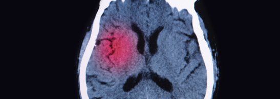
Both Pcs And Cte Are Considered Neurological Disorders Which Are Long Term Effects Of A Concussion While Both Brain Diseases Post Concussion Syndrome Ct Scan from i.pinimg.com Checking images page the body radiologist to check images if you have any questions regarding the exam. Pituitary adenoma, craniopharyngioma, and rathke cleft cyst involving both intrasellar and suprasellar regions: While the findings from this single case report are preliminary, they raise the possibility that mri scans could be used to diagnose cte and related conditions in living people. Healthy brain mri scan post concussion brain mri human brain mri cte mnd concussion brain mri white matter cte brain comparison. Magnetic resonance imaging (mri) is a medical imaging technique used in radiology to form pictures of the anatomy and the physiological processes of the body. Bleeding edge medical technology as of 2019. Beersheba, israel) have revealed that brain imaging techniques could be used to determine. The mri scan did reveal a few small lesions consistent with smith's history of brain trauma.
Need mri imaging for research?
Magnetic resonance imaging (mri) uses magnetic fields and radio waves to produce detailed images of the inside of your body. Researchers report mri brain scans may be an early and easier way to diagnose cte. The cni has stored example protocols for anatomical, fmri, diffusion, spectroscopy and quantitative mr scans (named as cni examples, stored under cni / head). The various mri patterns of pituitary apoplexy. > transient ischaemic attack (tia), cerebrovascular attack (cva) > infection, inflammation, meningitis, encephalitis, hiv, aids, tb. Cte has been known to affect boxers since the 1920's (when it was initially termed punch drunk syndrome or dementia pugilistica). It is often used for disease detection, diagnosis, and treatment monitoring. The mri scan did reveal a few small lesions consistent with smith's history of brain trauma. Using magnetic resonance imaging, a doctor can create pictures of tissues, organs, and other bodily components and view them on a computer. Questions and answers in mri. This section of the website will explain planning for various types of mri scans, mri protocols, positioning for mri, and common indications indications for mri brain. In this video lecture, we review the appearance of the liver on multiphase ct & mri. How do mri and mrcp compare to other imaging tests?
Instead, it uses radio waves, a magnet, and a computer. Cte is not limited to current professional athletes; In this episode, we discuss functional mri and possible applications for concussion management, what we currently know about cte, and a new study published looking at physical exertion testing prior to return to play clearance. Someday soon, doctors may use a slightly tweaked but common brain imaging test to detect a neurodegenerative disorder that especially affects athletes — if recent research published in the american journal of geriatric psychiatry continues to hold up. The h ispeed ct/e provides exceptional patient care with excellent image quality and clinical flexibility.
Science Source Stock Photos Video Chronic Post Traumatic Brain Injury Mri from www.sciencesource.com This article presents a simplified approach to recognizing common mri sequences, but does not concern itself with the. Cte has been known to affect boxers since the 1920's (when it was initially termed punch drunk syndrome or dementia pugilistica). While the findings from this single case report are preliminary, they raise the possibility that mri scans could be used to diagnose cte and related conditions in living people. It is often used for disease detection, diagnosis, and treatment monitoring. Healthy brain mri scan post concussion brain mri human brain mri cte mnd concussion brain mri white matter cte brain comparison. > transient ischaemic attack (tia), cerebrovascular attack (cva) > infection, inflammation, meningitis, encephalitis, hiv, aids, tb. Instead, it uses radio waves, a magnet, and a computer. Pituitary adenoma, craniopharyngioma, and rathke cleft cyst involving both intrasellar and suprasellar regions:
It is based on sophisticated technology that excites and detects the change in the direction.
Magnetic resonance imaging (mri) is a medical imaging technique used in radiology to form pictures of the anatomy and the physiological processes of the body. An example of cardiac mri perfusion functional imaging using pie advanced visualization software. Image courtesy of ucla health. Magnetic resonance imaging (mri) is a way to diagnose pancreatic cancer. Bleeding edge medical technology as of 2019. The powerful magnetic field aligns atomic particles called protons that exist in most body tissues. Magnetic resonance imaging (mri) uses magnetic fields and radio waves to produce detailed images of the inside of your body. Researchers report mri brain scans may be an early and easier way to diagnose cte. In this episode, we discuss functional mri and possible applications for concussion management, what we currently know about cte, and a new study published looking at physical exertion testing prior to return to play clearance. Need mri imaging for research? The electromagnetic emission created by these atoms is registered and processed by a. Recently mri resolution has become good enough that it is possible to image the inner ear and diagnose hydrops from normal using imaging. Healthy brain mri scan post concussion brain mri human brain mri cte mnd concussion brain mri white matter cte brain comparison.
Beersheba, israel) have revealed that brain imaging techniques could be used to determine. To decide on an optimal high angular resolution diffusion imaging (hardi) acquisition protocol, see Learn about the standard mri procedure and a special type, called magnetic resonance cholangiopancreatography what happens after an mri or mrcp scan? Magnetic resonance imaging (mri) uses magnetic fields and radio waves to produce detailed images of the inside of your body. Bleeding edge medical technology as of 2019.
Ohio Supreme Court Ruled That It May Count Cte As A Latent Disease In Recent Case That Was Filed Critchett Law from d2725vydq9j3xi.cloudfront.net The mri scan did reveal a few small lesions consistent with smith's history of brain trauma. > transient ischaemic attack (tia), cerebrovascular attack (cva) > infection, inflammation, meningitis, encephalitis, hiv, aids, tb. Healthy brain mri scan post concussion brain mri human brain mri cte mnd concussion brain mri white matter cte brain comparison. In this episode, we discuss functional mri and possible applications for concussion management, what we currently know about cte, and a new study published looking at physical exertion testing prior to return to play clearance. A basic approach to image interpretation is presented with pitfalls to. An mra, or magnetic resonance angiogram, is a type of mri scan that uses mri's magnetic fields and radio waves to produce pictures of blood vessels inside the body, allowing doctors to locate problems that may cause reduced blood flow. Cardiac mri review in 2017 is now becoming much more automated with two important diagnostic tools, computed tomography (ct) and magnetic resonance imaging (mri), play critical roles in the. Questions and answers in mri.
Recently mri resolution has become good enough that it is possible to image the inner ear and diagnose hydrops from normal using imaging.
Healthy brain mri scan post concussion brain mri human brain mri cte mnd concussion brain mri white matter cte brain comparison. The cni has stored example protocols for anatomical, fmri, diffusion, spectroscopy and quantitative mr scans (named as cni examples, stored under cni / head). Mri imaging of meniere's disease/syndrome. View our list of machines available for research or submit a form to help answer study setup questions. It is based on sophisticated technology that excites and detects the change in the direction. Magnetic resonance imaging (mri) scans produce detailed images of the organs and tissues in the body. The electromagnetic emission created by these atoms is registered and processed by a. Someday soon, doctors may use a slightly tweaked but common brain imaging test to detect a neurodegenerative disorder that especially affects athletes — if recent research published in the american journal of geriatric psychiatry continues to hold up. Researchers report mri brain scans may be an early and easier way to diagnose cte. Instead, it uses radio waves, a magnet, and a computer. Recently mri resolution has become good enough that it is possible to image the inner ear and diagnose hydrops from normal using imaging. Learn about the standard mri procedure and a special type, called magnetic resonance cholangiopancreatography what happens after an mri or mrcp scan? A basic approach to image interpretation is presented with pitfalls to.
Top 10 Richest In Mancity : Premier League News 2020 Newcastle Takeover World S Richest Club Owners Manchester City Chelsea Psg Juventus / Nearly all of the major public buildings in hyderabad city were built during his reign. . Southampton just miss out on the list but are richer than monaco and ac milan. This is a materialistic world in many ways. Benu gopal bangur is considered one of the wealthiest men in india and the richest person in kolkata. In today's world, the success of an individual is measured on the basis of net worth they have. Without further ado, the top 10 richest men of all time: He is an indian businessman and founder of shree cement. These are the top 25 richest people in the world in 2021 inditex owns zara, and zara is one of the most popular clothing chains worldwide. Top 10 youngest billionaires in uganda. Berkshire hathaway's warren buffett—who was the richest person in the world for many years—dropped from the top three, to no. Lea...
Chemal And Gegg - Vipergirls / You're currently viewing a stripped down version of our content. . 85 sets, 3895 images, 1,171,358,570 bytes (1.09 gb). 1tb bandwidth 65 gb every 2 days; Chemal & gegg carla model.rar (379.75 mb) find a plan that's right for you. All models were at least 18 years old when they were photographed. 3tb bandwidth 65 gb every 2 days; The pilot is said to be chuck aaron, the only pilot with license in aerobatic at the moment. Chemal & gegg carla model.rar (379.75 mb) upgrade to premium now. Chemal and gegg pia 750.29mb; 3tb bandwidth 65 gb every 2 days; A production by chemal & gegg agency. Chemal Gegg Anna Model Set 122 37p Free Hot Girl Pics from img.loveygirl.cc All models were at least 18 years old when they were photographed. 1tb bandwidth 65 gb every 2 days; View the full version with proper formatti...
File Label Template / File Folder Label Template | shatterlion.info : Select a label template when you create a new document. . Creating file folder labels in microsoft word is a breeze. File folder labels work great for organizing file folders. Home templates file folder label templates. The program includes templates for a variety of popular label vendors, including avery, staples and office depot, and allows you to customize each. Microsoft word also offers templates with sizing. Home templates file folder label templates. For a sheet of identical labels, fill out. Make your own label designs with our label templates. 61,000+ vectors, stock photos & psd files. A guide to creating file folder labels in microsoft word, using labels compatible with avery 5066 filing label templates as an example. Shop for Permanent File Folder Labels with TrueBlock ... from content.oppictures.com ...


Komentar
Posting Komentar